Back Muscles Anatomy Labeled / Diagrams of Back Muscles | 101 Diagrams / Ap1 lab 8 superficial muscles of the back and muscles of the arm.. Start studying 08 back muscles label. Learn anatomy back labeling with free interactive flashcards. The lats are attached to the upper end of the arm bones (humeri) at one end and fan out down the length of the spine to the pelvis. There is a dissection assistance pdf file that you can use to assist you in your lab preparation. Related posts of muscles labeled front and back muscle anatomy triceps.
Anatomynote.com found anatomy of back muscles diagram from plenty of anatomical pictures on the internet. Broadly considered, human muscle—like the muscles of all vertebrates—is often divided into striated muscle, smooth muscle, and cardiac muscle. For more anatomy content please follow us and visit our website: We hope this picture anatomy of back muscles diagram can help you study and research. Anatomy of the back muscles.
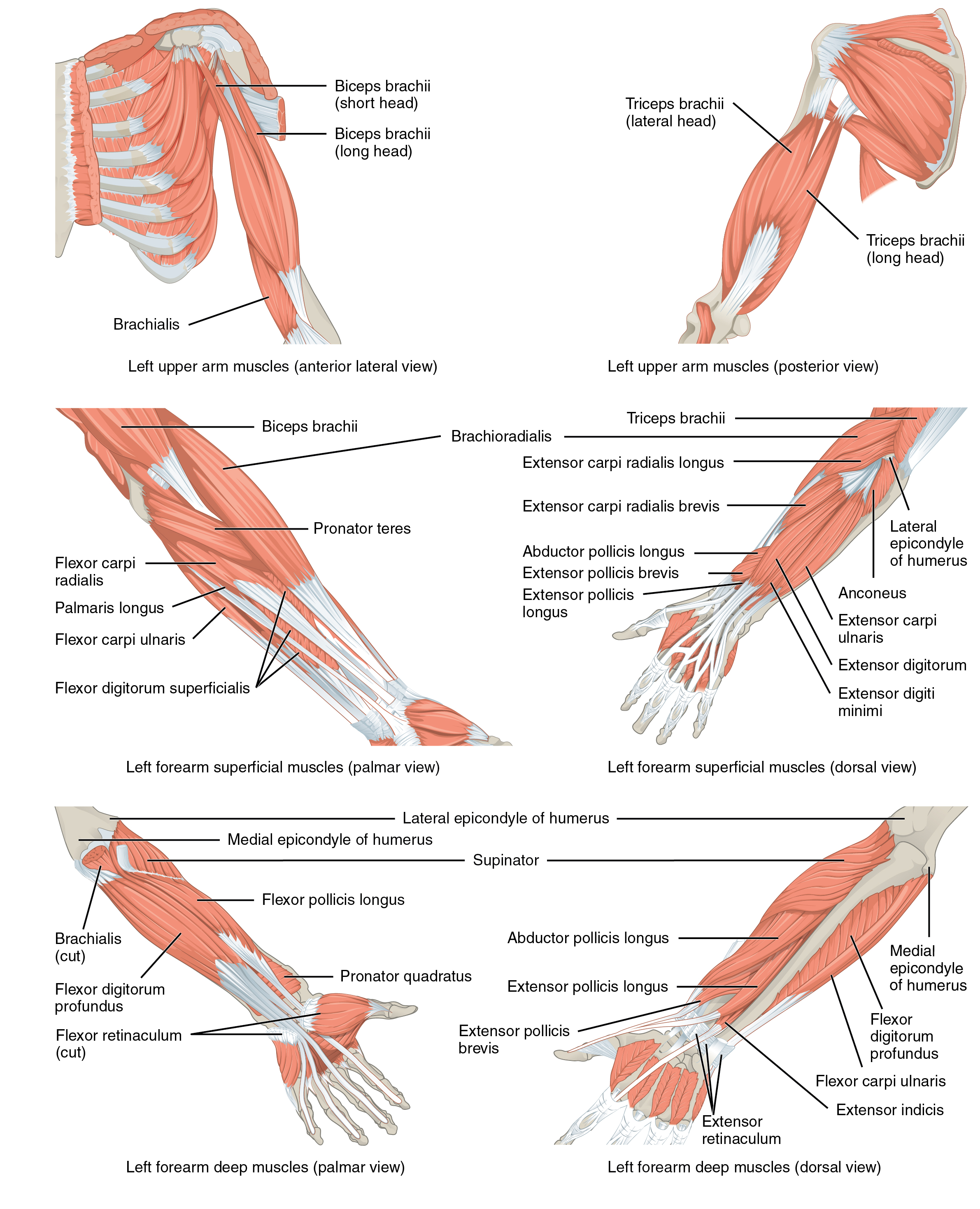
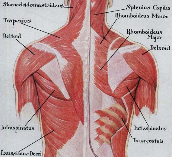
Muscles of the Pectoral Girdle and Upper Limbs · Anatomy ... from philschatz.com The back anatomy includes the latissimus dorsi, trapezius, erector spinae, rhomboid, and the teres major. Browse 3,565 back anatomy muscles stock photos and images available, or start a new search to explore more stock photos and images. Superficial back muscles, intermediate back muscles and intrinsic back muscles.the intrinsic muscles are named as such because their embryological development begins in the back, oppose to the superficial and intermediate back muscles which develop elsewhere and are therefore classed as extrinsic muscles. All activity in the body is regulated by muscle mass. Muscle mass are just how we move as well as live. Human muscle system, the muscles of the human body that work the skeletal system, that are under voluntary control, and that are concerned with movement, posture, and balance. We think this is the most useful anatomy picture that you need. During a chin up the lats serve to bring the body up towards the arm.
The deltoid, teres major, teres minor, infraspinatus, supraspinatus (not shown) and subscapularis muscles (not shown) all extend from the scapula to the humerus and act on the shoulder joint.
In this image, you will find 1st cervical vertebrae, atlus, cervical plexus, 7th cervical vertebrae, 1st thoracic vertebrae, brachial plexus, spinal dura mater, filaments of spinal nerve roots, 12th thoracic vertebra, 1st lumber vertebra, iliohypogastric nerve, ilioinguinal nerve, lumbar. Choose from 500 different sets of anatomy back labeling flashcards on quizlet. Browse 3,579 back muscle anatomy stock photos and images available, or search for pelvic bone or lymphatic system to find more great stock photos and pictures. Sale human anatomy vintage chart of body front back skeleton and muscle system bone mass graphic single shower curtain, major muscles on the back of the body, amazon com exercise and muscle guide female fitness, muscles german names chart muscular body, the nervous system chart rear. We think this is the most useful anatomy picture that you need. The shoulder muscles are responsible for maintaining the widest range of motion of any joint in your body. These muscles include the large paired muscles in the lower back called erector spinae which help hold up the spine and gluteal muscles. Browse 3,565 back anatomy muscles stock photos and images available, or start a new search to explore more stock photos and images. These important muscles control many motions that involve moving the arms and head — such as throwing a ball, looking up at the sky, and raising your hand. Superficial back muscles, intermediate back muscles and intrinsic back muscles.the intrinsic muscles are named as such because their embryological development begins in the back, oppose to the superficial and intermediate back muscles which develop elsewhere and are therefore classed as extrinsic muscles. Labeled anatomy chart of male triceps and back muscles on white background labeled human anatomy diagram of man's arm, shoulder and upper back muscles in a posterior view on a white background. These structures work together to support the body, enable a range of movements, and send messages from the brain to the. Start studying 08 back muscles label.
All activity in the body is regulated by muscle mass. See back muscle anatomy stock video clips. Trapezius muscles are the site of many a sore spot. Human musculature bodybuilding infographic muscular system vector human anatomy back muscle anatomy bicep male muscular anatomy human body anatomy female female anatomy muscle hamstrings muscle. Back muscle diagrams labeled, lower back muscle diagrams labeled, human muscles, back muscle diagrams labeled, lower back muscle diagrams labeled.
Muscles Back Posterior Human Anatomy Vintage Medical Chart from img.etsystatic.com For more anatomy content please follow us and visit our website: The extrinsic muscles that are associated with upper extremity and shoulder movement and the intrinsic muscles that deal with movements of the vertebral column. The back anatomy includes some of the most massive and functionally important muscles in the human body. Both the deltoid and the trapezius are firmly attached to the spine of the scapula. Sale human anatomy vintage chart of body front back skeleton and muscle system bone mass graphic single shower curtain, major muscles on the back of the body, amazon com exercise and muscle guide female fitness, muscles german names chart muscular body, the nervous system chart rear. Muscle mass are just how we move as well as live. Superficial back muscles, intermediate back muscles and intrinsic back muscles.the intrinsic muscles are named as such because their embryological development begins in the back, oppose to the superficial and intermediate back muscles which develop elsewhere and are therefore classed as extrinsic muscles. Browse 3,565 back anatomy muscles stock photos and images available, or start a new search to explore more stock photos and images.
Some muscle mass specifically 7 deep muscles of back anatomy work without us assuming, like our heart whipping, while other muscle mass are regulated by our thoughts as well as permit us to do stuff as well as walk around.
Back muscle diagrams labeled, lower back muscle diagrams labeled, human muscles, back muscle diagrams labeled, lower back muscle diagrams labeled. Some of these muscles are quite large and cover broad areas. During a chin up the lats serve to bring the body up towards the arm. Muscle mass are just how we move as well as live. Anatomy of the spine and back spine muscles diagram. The back's muscles start at the top of the back (named the cervical vertebrae) and go to the tailbone (also named the coccyx). The back is the body region between the neck and the gluteal regions. Mastoid process and lateral end of the superior nuchal line: Anatomy of back muscles your back consists of three distinct layers of muscles, namely the superficial layer, the intermediate layer, and the deep layer. On this page, you'll learn about each of these muscles, their locations and functional anatomy. These important muscles control many motions that involve moving the arms and head — such as throwing a ball, looking up at the sky, and raising your hand. Start studying 08 back muscles label. These muscles include the large paired muscles in the lower back called erector spinae which help hold up the spine and gluteal muscles.
Out of these, the cookies that are categorized as necessary are stored on your browser as they are essential for the working of basic functionalities of the website. See back muscle anatomy stock video clips. Human musculature bodybuilding infographic muscular system vector human anatomy back muscle anatomy bicep male muscular anatomy human body anatomy female female anatomy muscle hamstrings muscle. Ap1 lab 8 superficial muscles of the back and muscles of the arm. The back functions are many, such as to house and protect the spinal cord, hold the body and head upright, and adjust the movements of the upper and lower limbs.
he Muscular System - Deep Layers, Back Laminated Anatomy Chart from www.anatomy.link This website uses cookies to improve your experience while you navigate through the website. Learn anatomy back labeling with free interactive flashcards. Both the deltoid and the trapezius are firmly attached to the spine of the scapula. Back dissection, cranial neve 11, dissection, latisumus dorsi, lavator. #back muscle diagrams labeled #lower back muscle diagrams labeled. Labeled anatomy chart of male triceps and back muscles on white background labeled human anatomy diagram of man's arm, shoulder and upper back muscles in a posterior view on a white background. We think this is the most useful anatomy picture that you need. 12 photos of the back muscle diagrams labeled.
Browse 3,579 back muscle anatomy stock photos and images available, or search for pelvic bone or lymphatic system to find more great stock photos and pictures.
In this image, you will find 1st cervical vertebrae, atlus, cervical plexus, 7th cervical vertebrae, 1st thoracic vertebrae, brachial plexus, spinal dura mater, filaments of spinal nerve roots, 12th thoracic vertebra, 1st lumber vertebra, iliohypogastric nerve, ilioinguinal nerve, lumbar. The back anatomy includes the latissimus dorsi, trapezius, erector spinae, rhomboid, and the teres major. Broadly considered, human muscle—like the muscles of all vertebrates—is often divided into striated muscle, smooth muscle, and cardiac muscle. Anatomy of back muscles your back consists of three distinct layers of muscles, namely the superficial layer, the intermediate layer, and the deep layer. The muscles of the back can be arranged into 3 categories based on their location: Superficial back muscles, intermediate back muscles and intrinsic back muscles.the intrinsic muscles are named as such because their embryological development begins in the back, oppose to the superficial and intermediate back muscles which develop elsewhere and are therefore classed as extrinsic muscles. #back muscle diagrams labeled #lower back muscle diagrams labeled. The muscles of the chest and upper back occupy the thoracic region of the body inferior to the neck and superior to the abdominal region and include the muscles of the shoulders. Anatomy of the back muscles. Choose from 500 different sets of anatomy back labeling flashcards on quizlet. He is an attending emergency medicine phys. We think this is the most useful anatomy picture that you need. Human muscle system, the muscles of the human body that work the skeletal system, that are under voluntary control, and that are concerned with movement, posture, and balance.
Muscle anatomy triceps 12 photos of the muscle anatomy triceps anatomy of triceps muscle, biceps triceps muscle anatomy, muscle anatomy triceps, triceps muscle anatomy mri, human muscles, anatomy of triceps muscle, biceps triceps muscle anatomy, muscle anatomy triceps, triceps muscle anatomy mri back muscles anatomy. Choose from 500 different sets of anatomy back labeling flashcards on quizlet.


0 Komentar